|
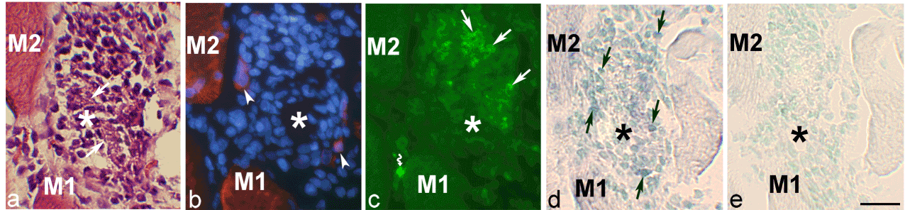
| Figure 6: Sections from a series of sections of a specimen from the 6w group, experimental side. The sections were stained with H&E (a), for demonstration of desmin (including DAPI reaction) (b), NK-1R immunolabelling (c) and for demonstration of TNF-alpha mRNA (in situ hybridization) (antisense staining in d, sense staining in e). Asterisks mark a previously existing (necrotic) muscle fiber, in the area of which there is an accumulation of white blood cells [arrows in (a) point at debris material of the fiber]. There is no desmin immunoreaction within the fiber, whilst there are desmin reactions in the most peripheral cells, which based on their strong desmin immunoreaction and occurrence of large nuclei are interpreted to be very small regenerating muscle fibers. There are cellular NK-1R immunoreactions in the necrotic muscle fiber (arrows). Artefact at curved arrow. Infiltrated cells do show TNF-alpha mRNA reactions (arrows in d). M1 and M2=corresponding muscle fibers. Bar=25 μm. |