|
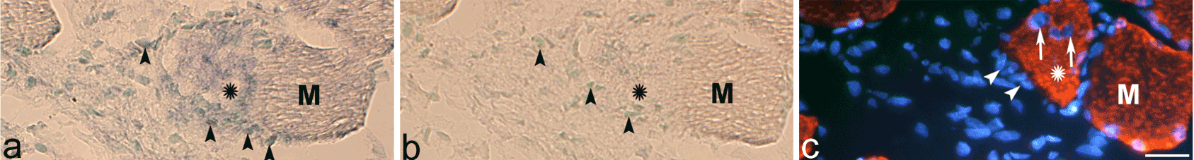
| Figure 9: Parallel sections processed for TNF-alpha mRNA (antisense staining in a, sense staining in b) and desmin immunoreaction (c) (DAPI in mounting medium). The specimen was from the 6w group, experimental side. One of the muscle fibers (asterisk) shows a very strong non-striated desmin immunoreaction. There are intracellular nuclei (arrows, c) and patchy TNF-alpha mRNA reactions in this muscle fiber. There are also TNF-alpha mRNA reactions in cells located just outside this muscle fiber (arrowheads). There are no reactions in the sense control section (b). M=corresponding muscle fiber. Bar=25 μm. |