|
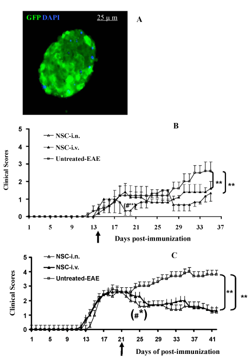
| Figure 1: Effective suppression of EAE by i.n. administration of GFPaNSCs. (A) A modified neurosphere is shown. GFP+ cells were sorted by FACS to reach >99% purity, then transferred into growth medium where they became neurospheres. Nuclei in A are stained with DAPI (blue). Magnification ×40. aNSCs transduced with Lv.GFP were dissociated into single cells and washed twice with PBS. 1.0×10⁶ cells/mouse were injected once, at different stages of disease, EAE mice that received the same volume of PBS either i.n. or i.v. served as a sham injection control. Symbols represent mean values and SD of 6-7 mice from each group. (B) NSC-i.n. delivery or NSC-i.v. injection at day 14 p.i.; (C) NSC-i.n. delivery or i.v. injection at the peak of disease (day 21 p.i.). *p<0.05, **p<0.01, comparisons between sham-EAE group and other groups; #p<0.05, comparison between NSC-i.n. and NSC-i.v. injection. |