|
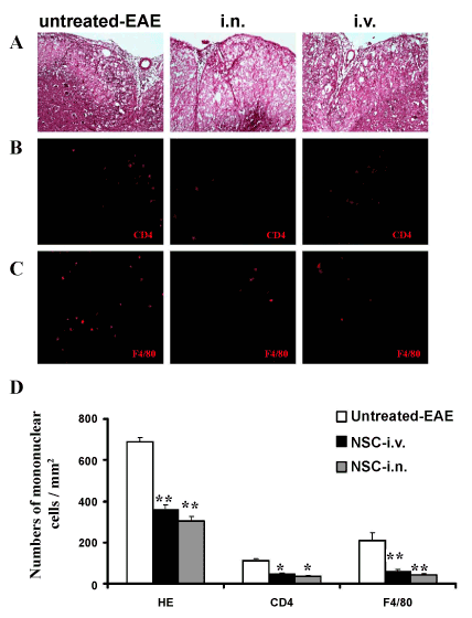
| Figure 3: Anti-inflammatory effect of aNSCs in the CNS. Mice described in Figure 1 were sacrificed 21 days p.t., and lumbar spinal cords were harvested for H&E staining and immunostaining. All groups were examined in the same region of the ventral column at L3. Reduced inflammatory infiltration (A) and numbers of CD4+ cells (B) and F4/80+ cells in (C) were found in spinal cord lesions of i.n. delivery and i.v. injection mice compared to untreated EAE mice. Magnification ×20 for A, B and C. (D) Quantitative analysis of H&E scores and numbers of CNS-infiltrating mononuclear cells. Symbols represent mean values and SD of 6-8 mice from each group. *p<0.05, **p<0.01, comparison between untreated-EAE group and other groups. There was no significant difference between the i.n. delivery and i.v. injection groups. |