|
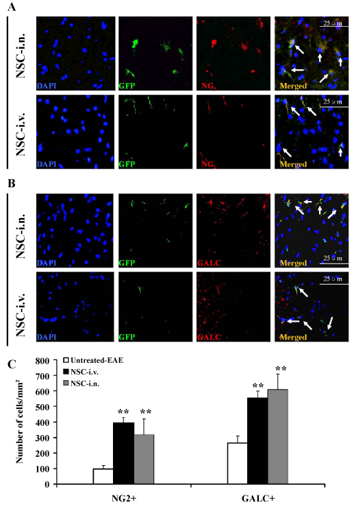
| Figure 6: I.n.-GFP-aNSCs promote remyelination of demyelinated axons in spinal cord. Spinal cords were harvested for immunohistology at day 21 p.t. All groups were examined in the same region: a specific site on the ventral column of the lumbar spinal cord (L3). Cells co-labeled with GFP (green), cell nucleus (blue) and oligodendrocyte precursor cells (NG2+; red) (A) and oligodendrocytes (GalC+; red) (B) derived from transplanted aNSCs (arrows). (C) Quantification of total NG2+GFP+ and GalC+GFP+ cell numbers. **p<0.01, comparison between untreated-EAE and other groups. Symbols represent mean values and SD of 6-8 mice from each group. Magnification ×40 for A-B. |