|
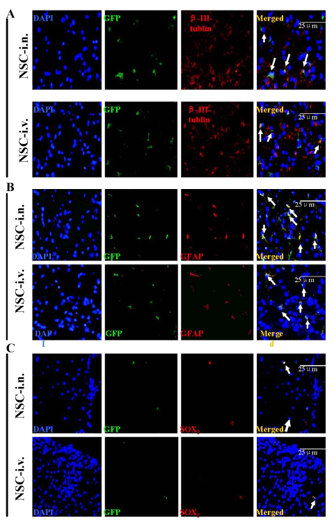
| Figure 7: I.n.-GFP-aNSCs selectively expand neuron and oligodendrocyte populations in vivo. Mice treated with aNSCs at day 21 p.i. were sacrificed at day 21 p.t., and lumbar spinal cords were harvested for immunohistology. Cells co-labeled with GFP (green), cell nucleus (blue) and neural specific markers (red), including β–III-tublin (A) GFAP+ (B) were identified as differentiated cells derived from transplanted GFP-aNSCs (arrows). Some transplanted GFPaNSCs remained SOX2+ (C) undifferentiated NSCs. Magnification ×40. |