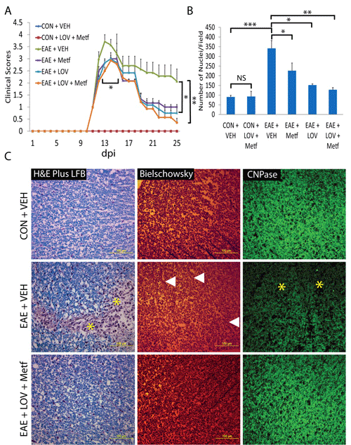
EAE was induced with guinea pig MBP (25 μg/rat) in female Lewis rats and treatment of LOV (1 mg/kg) and/or Metf (150 mg/kg) or vehicle were started orally by gavages in rats with established EAE or in healthy controls (CON). A: Shown is the composite mean ± SEM of clinical scores in rats (n=9)/group evaluated in three separate experiments. B: Shown is the composite mean ± SEM of three to four samples/group analyzed for cellular infiltration in the SCs of EAE rats on peak clinical day. C: Representative photographs of the cross-section of lateral funiculi of the SCs of rats (n=6)/group on peak clinical day, stained with LFB plus H&E (left panel) depicting cellular infiltration and demyelination, bielschowsky staining (middle panel) depicting axonal integrity and immunostaining with anti-CNPase (right panel) depicts demyelination. Asterisks depict cellular infiltration and demyelination in the white mater of the SC. Triangles depict neuronal axonal loss in the white matte of the SC. Differences were statistically significant as indicated: *p<0.05, **p<0.01 and ***p<0.001, and NS (not significant).