 |
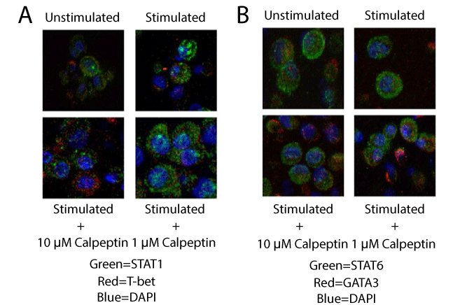
| Figure 6: Immunofluorescent staining for localization of transcription factor. Representative images of MBP Ac1-11-specific T cells stimulated with MBP Ac1-8 peptide for 48 hours and then stained with immunofluorescent staining for: A. STAT1 and T-bet and B. STAT6 and GATA3. The nucleus of the T cells was stained with DAPI. In both staining paradigms the calpeptin treated groups demonstrated a larger percentage of cells staining positive for the transcription factors T-bet and GATA3 when compared with untreated control and untreated stimulated group (n=3 for all treatments and staining groups). |