|
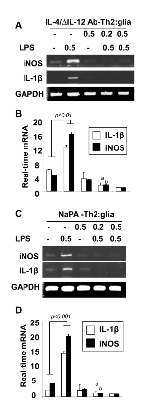
| Figure 2: Pre-conditioned MBP-primed Th2 cells suppress iNOS and IL- 1β in mouse primary microglia. MBP-primed T cells from SJL/J mice were treated with IL-4 plus anti-IL-12 antibody combination (IL-4/ΔIL-12 Ab) and NapA for 4 days to polarize Th2 condition followed by the treatment on LPS-stimulated mouse primary microglia. After 5 hrs of incubation with IL-4/ΔIL-12 Ab, the mRNA expression of iNOS and IL-1β were analyzed by RT-PCR (A) and realtime PCR analysis (B). ap<0.001 vs. LPS-stimulated IL-1β; bp<0.001 vs. LPS-stimulated iNOS. After 5 hrs of incubation with NaPA, the mRNA expression of iNOS and IL-1β were analyzed by RT-PCR (C) and realtime PCR analysis (D). ap<0.001 vs. LPS-stimulated IL-1β; bp<0.001 vs. LPS-stimulated iNOS. Data are mean ± SD of three independent experiments. |