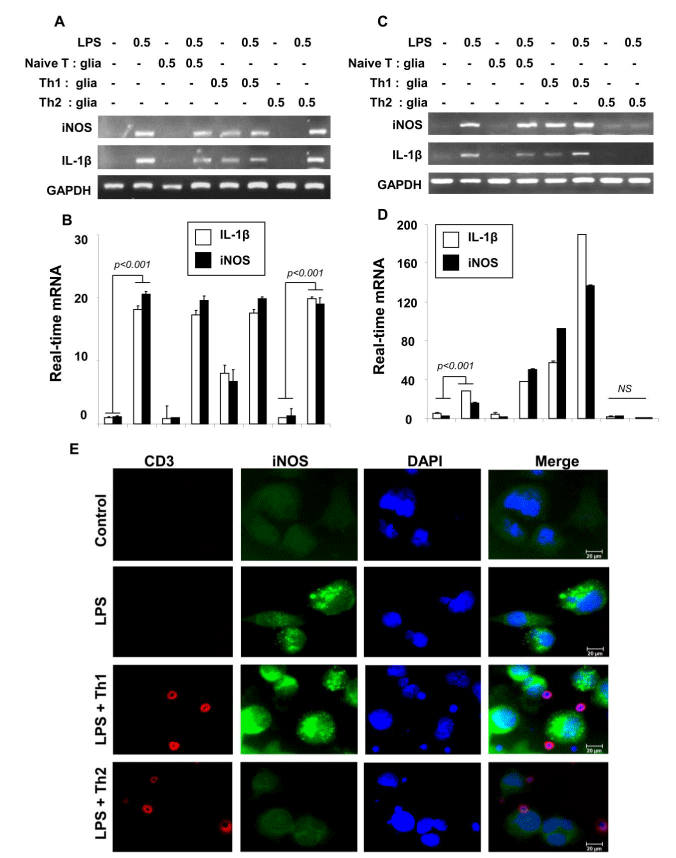
Microglia received different concentrations of Th2 cells within insert. After 1 hr, inserts are removed followed by the stimulation of microglial cells with LPS (0.5 μg/ ml) for 1 hr in serum-free condition. After another 5 h of incubation, the mRNA expression of iNOS and IL-1β was analyzed by RT-PCR (A) and by real-time PCR (B). Next, Microglia were stimulated by plasma membranes (equivalent to 0.5:1 of T cell: glia) of normal T cells, MBP-primed Th1 and MBP-primed Th2 cells separately. After 1 hr, microglial cells were stimulated with LPS (0.5μg/ml) LPS. After another 5 h, microglial cells were analyzed for the expression of iNOS and IL-1β mRNAs by semi-quantitative RT-PCR (C) and quantitative real-time PCR (D). (E) Immunocytochemical analyses showed the interaction between CD3-immunostained T cells (red) and iNOS (green) positive LPS-treated microglia. Data are the mean ± S.D. of three different experiments. NS=not significant.