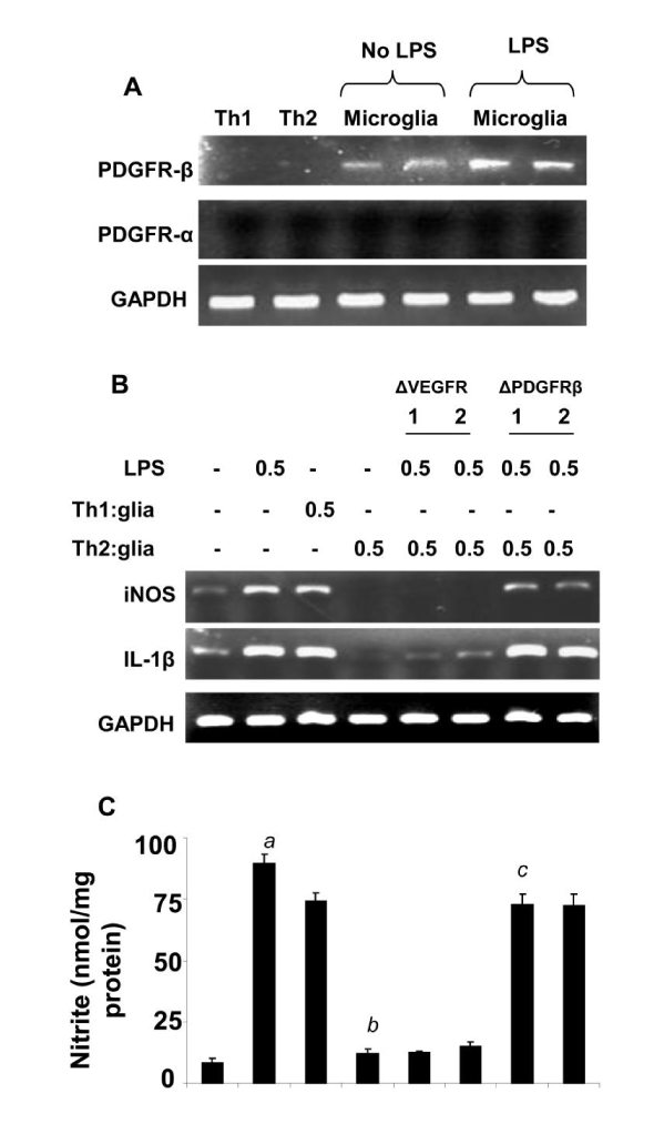
(A) T cells (MBP-primed Th1 and Th2) and primary microglia (with or without LPS) were analyzed for PDGFRα and PDGFRβ mRNAs by semi-quantitative RT-PCR. Microglia were incubated with different concentrations of neutralizing antibodies against either PDGFRβ or VEGF-R (1-2 μg/mL). Excess antibodies were removed after 1 h of incubation followed by the stimulation of microglia by Th2 cells at a ratio of 0.5:1 T cell: microglia. After 1 h of stimulation, T cells were removed followed by incubation of adherent microglia with LPS (0.5 μg/ ml) in serum-free media. After another 5 h of incubation, the expression of iNOS and IL-1β was analyzed by RT-PCR (B). After 24 h of incubation (total), supernatants were used to assay nitrite by Griess method described under “Materials and Methods” section (C). Data are mean ± S.D. of three different experiments. ap<0.001 vs. control , bp<0.001 vs. Th1-treated ,and cp<0.001 vs. Th2-treated microglial cells.