|
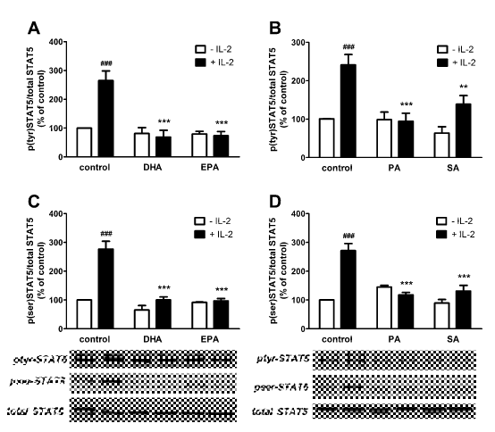
| Figure 3: Effects of 50 μM DHA, EPA, SA, and PA on IL-2-induced signal transducer and activator of transcription (STAT) 5 tyrosine 694 and serine 726 phosphorylation. Lymphocytes were incubated with 5 μg/mL ConA for 24 h. Afterwards, lymphocytes were washed with PBS and cultured with the different fatty acids in the presence or absence of IL-2 (30 ng/mL) for 1 h. Total proteins were extracted from lymphocytes for western-blotting analysis. Blots were analyzed by densitometry and the results normalized to their respective controls, which were set to a value of 100% for each experiment. The values are presented as the means ± SEM. ###p<0.001 for comparison with the control in the absence of IL-2); **p<0.01 and ***p<0.001 for comparison with the control treated with IL-2. |