 |
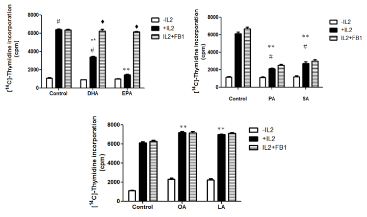
| Figure 9: Effect of 50 μM DHA, EPA, SA, and PA and 25 μM OA and LA on IL2 induced lymphocyte proliferation in the presence and absence of fumonisin B1 (FB1). Lymphocytes were stimulated with ConA for 24 h, washed, and incubated with 10 μM FB1, 30 ng/mL IL-2 and 50 μM DHA, EPA, SA, and PA and 25 μM OA and LA for 48h. Data are expressed as counts per minute (cpm) and presented as mean ± S.E.M. of three determinations from three experiments. The values are presented as mean ± S.E.M. of four determinations from three experiments. #p<0.05 for comparison between fatty acid treatments vs. the control (in the absence of FAs and IL-2); **p<0.05 for comparison between fatty acid treatments vs. the control treated with IL-2; ♦p<0.05 for comparison between fatty acid treatments in the presence of wortmannin vs. fatty acid treatments in the presence of IL-2. |