|
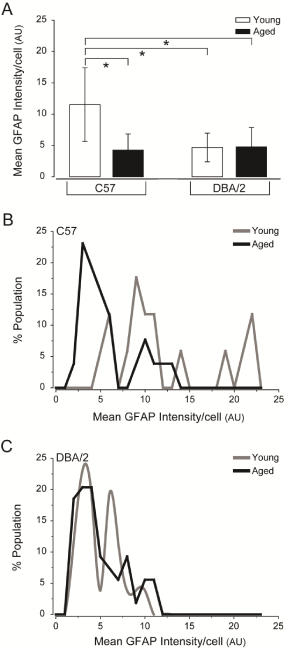
| Figure 4: Decreased GFAP expression/cell is a common characteristic of retina challenged by glaucoma-related stressors. (A) Quantification of the mean GFAP intensity/cell (arbitrary units; y-axis) across all images and samples reveals that mean GFAP intensity/cell is higher in young C57 retina than in young DBA/2, aged C57 and aged DBA/2 retina. Error bars represent ± standard deviation and asterisks indicate p<0.05. (B) Histogram function of GFAP intensity/cell for young C57 (gray) and aged C57 (black) retina plotted as percent population (y-axis) versus GFAP intensity/cell (arbitrary units, x-axis). The histogram function for aged C57 retina reveals a shift towards lower GFAP intensity and a smaller range of distribution, as compared to young C57 retina. (C) Histogram function of GFAP intensity/ cell for young DBA/2 (gray) and aged DBA/2 (black) retina plotted as percent population (y-axis) versus GFAP intensity/cell (arbitrary units; x-axis). The histogram function for young DBA/2 and aged DBA/2 retina demonstrate a similar range of distributions that is shifted towards lower GFAP expression. However, young DBA/2 retina exhibits the presence of two distinct peaks of GFAP expression that account for almost 50% of GFAP expression in the population, while aged DBA/2 retina exhibits a single peak accounting for only 20% of astrocyte population. |