|
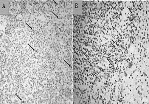
| Figure 2: Immunostainning of p24 antigen in tissue (A) Distribution of p24 antigen is observed as dark brown stained cells infiltrating the surrounding tissue (indicated by arrows). (B) The distribution of CD56+ cells is seen throughout the tissue as brown stained cells. |