|
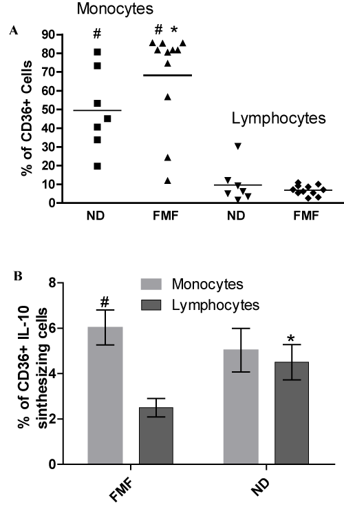
| Figure 1: Flow cytomety analysis of CD36+ monocytes and lymphocytes (A) and CD36+ cells, intracellularly synthesizing IL-10 (B) in ND and FMF patients. PBMC were surface stained by anti-CD36, CD3 or CD14 and fixed and permeabilized cells incubated with anti-human IL-10 or matched isotype control and subjected to FACS analysis. All data represent means ± SEM (error bars) and are significantly different comparing FMF with ND (* Pw<0.05) or comparing monocytes with lymphocytes (# Pt<0.001). |