 |
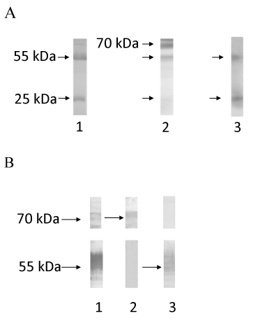
| Figure 2: A. SDS-PAGE (10% acrylamide) of purified human IgG (lane 1), CA215 (lane 2) and CIgG (lane 3). The molecular weights of protein bands are indicated by arrows as 70 kDa, 55 kDa or 25 kDa, respectively, for immunoglobulin heavy chain (IgM and IgG) and light chains, respectively. B. Western blot assay of CA215 to indicate protein bands probed with RP215 followed by ALP-labeled goat anti-mouse IgG (lane 1), or with ALP-labeled anti-human IgM (lane 2) and ALP-labeled anti-human IgG (lane 3), to indicate the position of human IgM and IgG heavy chains, respectively (described in text). |