|
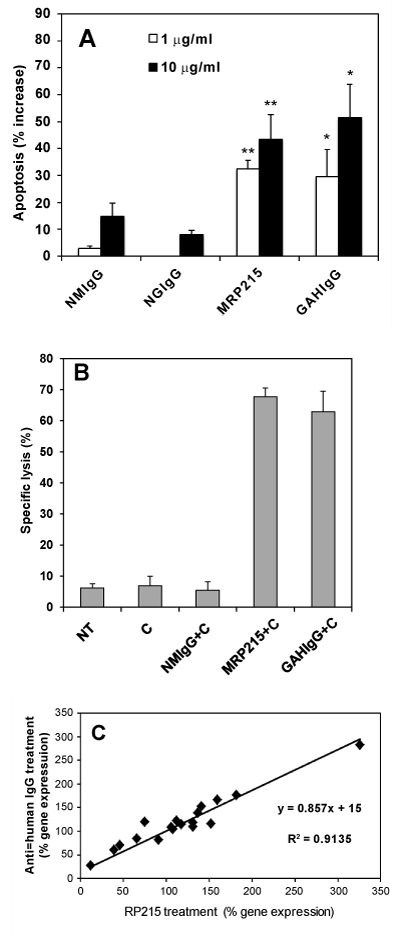
| Figure 4: A. Effects of treatment of different immunoglobulins on induced apoptosis of cultured OC-3-VGH cancer cells, including normal mouse IgG (NMIgG), normal goat IgG (NGIgG), mouse RP215 (MRP215), goat anti-human IgG (GAHIgG) Open and solid columns represent antibody concentrations of 1 μg/ml and 10 μg/ml, respectively. All data presented are statistically significant at * P<0.01 and ** P<0.001. B. Complement-dependent cytotoxicity (CDC) reaction in the presence or absence of complement and/or antibodies containing 10 μg/ml of mRP215 or normal immunoglobulins for the assays of cancer cells. Lane 1: no treatment (NT); Lane 2: freshly prepared rabbit baby complement (C) only (3 μl in each experiment); Lane 3: normal mouse IgG plus complement (NMIgG+C); Lane 4: mouse RP215 plus complement (MRP215+C); Lane 5: goat anti-human IgG plus complement (GAHIgG+C). C. Correlation analysis of RP215 and anti-human IgG on gene regulation changes to cancer cells in culture (OC-3- VGH and C-33A cancer cell lines). Each point (♦) represents for one gene used in the semi-quantitative RT-PCR experiment, including: IgG, NFκB-1, P0, P1, P2, c-fos, P21, cyclin D1, TLR-2, TLR-3, TLR-4, TLR-6, TLR-7 and TLR-9. A correlation coefficient of R2 = 0.9135 was obtained statistically. Details of these studies were presented previously in ref [9] (obtained with permission). |