 |
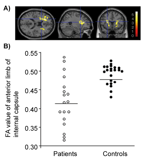
| Figure 2: White matter fractional anisotropy (FA) in stroke patients and control subjects. (A) Voxel-based analysis was performed using SPM8 software. FA maps of patients (n=18) and healthy subjects (n=22) were compared using analysis of covariance (ANCOVA), with age and sex as covariates. Statistical inferences were made with a voxel-level threshold of p< 0.001, uncorrected, and a minimum cluster size of 100 voxels. Statistical parametric mapping projections were superimposed on a representative magnetic resonance image (x=-26, y=12, z=18). FA in the right and left anterior limbs of the internal capsule was reduced in stroke patients. (B) Scatter plots of FA values in the regions of FA reduction in stroke patients. The regional FA value was calculated by averaging the FA values for all voxels within the volume of interest corresponding to the cluster composed of significant contiguous voxels in the preceding analysis. In the bilateral anterior limbs of the internal capsule, the FA values of stroke patients were lower than those of healthy subjects (p < 0.01). |