 |
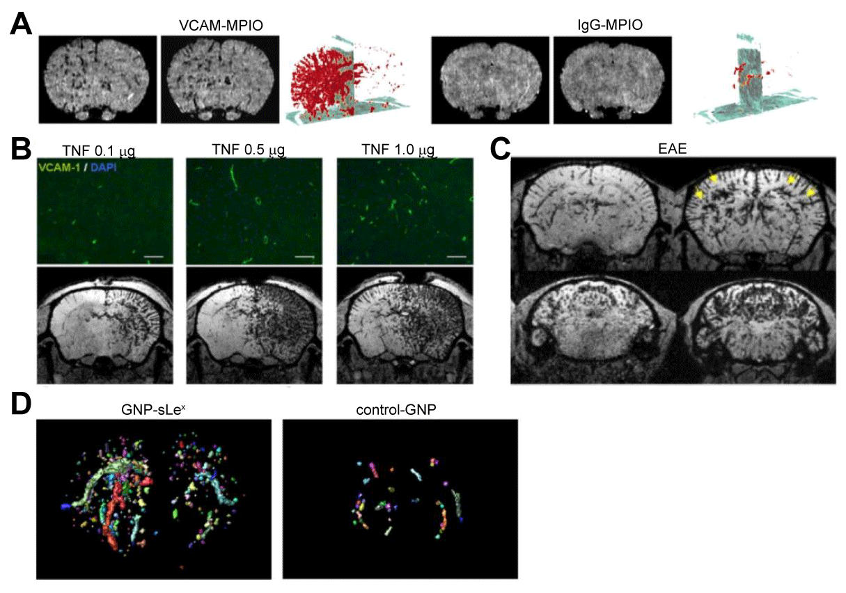
| Figure 3: MR Imaging of adhesion molecules in neuroinflammation. (A) VCAM-MPIO compared to IgG-MPIO enhanced MR imaging after intracerebral injection of IL-1β, axial T2*-weighted images and 3-dimensional volumetric maps of VCAM-MPIO (or IgG-MPIO) binding are shown. (B) VCAM-upregulation after intracerebral injection of TNF on histology and VCAM-MPIO-enhanced MRI. (C) Diffuse VCAM upregulation in the brain of an EAE mouse on VCAM-MPIO-enhanced MRI. (D) 3D reconstruction maps of GNP-sLex- and control GNPenhanced MRI in EAE mice reveal increased selectin expression in the inflamed brain. (Modified from McAteer etl al. [14], Montagne et al. [19], and Serres et al. [29] with permission). |