 |
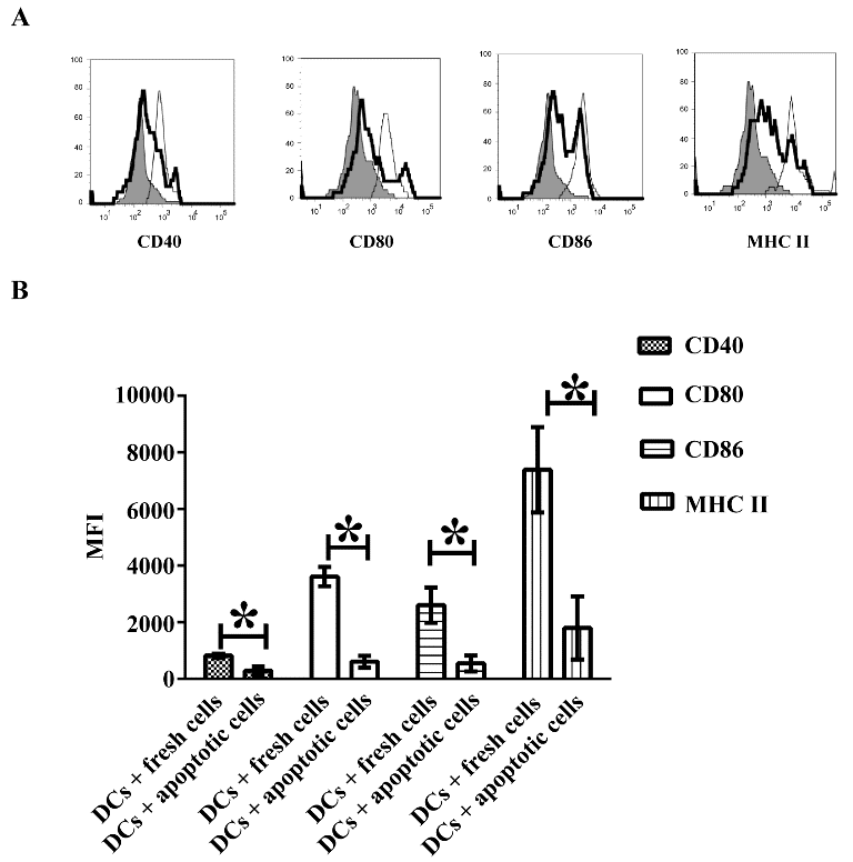
| Figure 1: Treatment of apoptotic T cells inhibits expression of multiple signal molecules on DCs. CD11c+ DCs were isolated from C57 BL/6J mice and sorted using flow cytometry. CD11c+ DCs were co-cultured with apoptotic (thick line) or fresh T cells (thin line) as a control. Cells were then washed and harvested. Expression of CD40, CD80, CD86 and MHC II was detected using flow cytometry. Isotype control is indicated by shading. Error bars shown in this figure represent mean and SD of triplicate determinations of MFI for protein expression on DCs in three independent experiments (n=3, t test, *P<0.05). |