 |
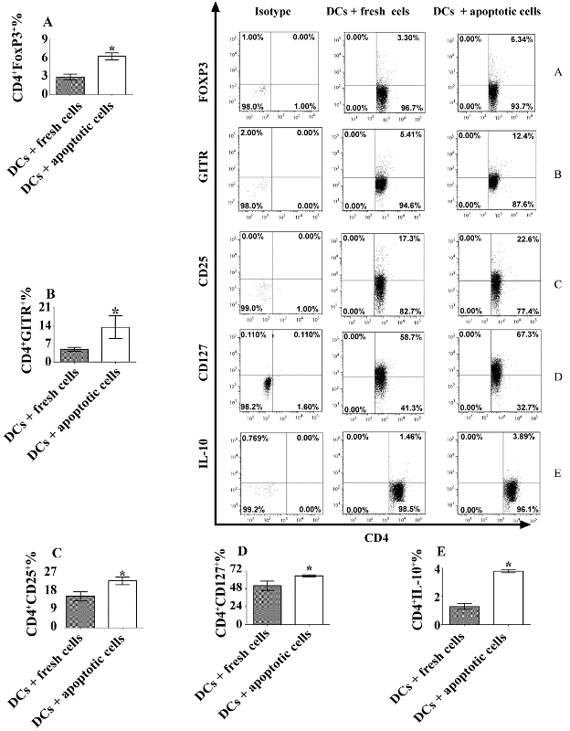
| Figure 4: Tolerogenic DCs induced by apoptotic T cells facilitate development of CD4+ regulatory T cells. CD11c+ DCs were isolated from C57BL/6J mice and sorted using flow cytometry. CD11c+ DCs were co-cultured at 37°C overnight with apoptotic or fresh T cells as a control. MOG specific CD4+CD25- T cells were isolated from 2D2 transgenic mice and incubated with 0.1 μM MOG peptide-pulsed DCs treated with apoptotic or fresh T cells as a control for 3 days at 37°C. Cells were then harvested and a flow cytometry assay was carried out. Expression of FoxP3 (A), GITR (B), CD25 (C), CD127 (D) and IL-10 (E) is shown. Isotype controls are also indicated. Error bars represent mean and SD of triplicate determinations of percentage (%) of expression of Treg-associated markers on CD4+ T cells in three independent experiments (n=3, t test, *P< 0.05). |