|
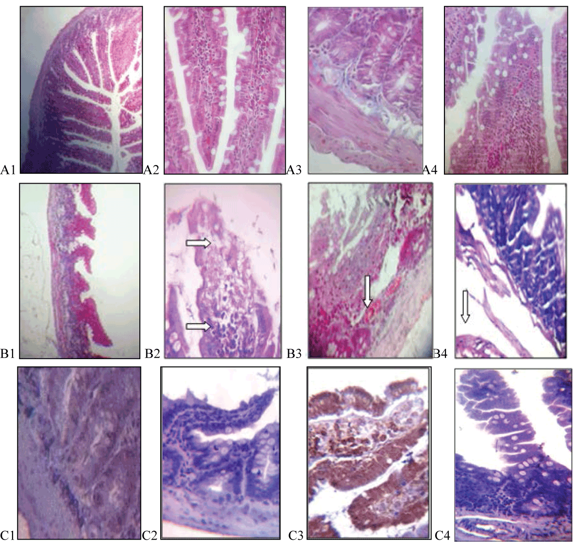
| Figure 2: Saline/chamomile e×tract treated group: On day 8, the jejunum shows nearly normal shape of villi (A1, Trichrome ×100), most of the villi seen cover by columnar epithelium, the crypt and the muscular layers appear normal (A2,A3 Trichrome ×400), or sometimes the epithelium of villi are missing with cellular infiltration in the lamina propria (A4, Trichrome ×400). On day 12, the villi appear with abnormal shape (B1, Trichrome ×40), the tips of some villi shows necrosis (upper arrow) with inflammatory cell infiltration in the lamina propria, lower arrow (B2,H&E ×400). Congested blood vessels in the lamina propria, arrow (B3, Trichrome ×400) with muscular edema are also seen, arrow (B4,H&E ×400). Mild and negative Ki-67 immuno reactivity in nuclei of cells at day eight and twelve respectively (C1, immunohistochemistry ×400, C2, immunohistochemistry ×400). Moderate and negative cytoplasmic reaction to Bcl-2 cells at day eight and twelve respectively (C3 immunohistochemistry ×400, C4, immunohistochemistry ×400). |