 |
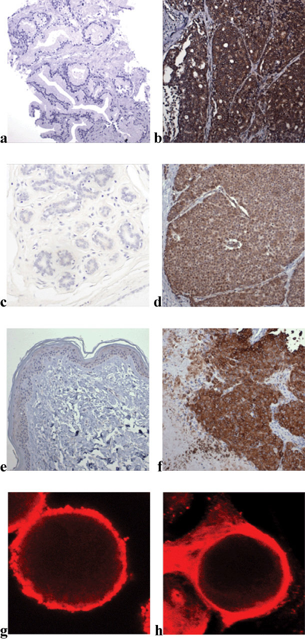
| Figure 1: Sections of: (a) normal prostate; (b) prostate cancer; (c) normal breast; (d) invasive breast cancer; (e) normal skin; and (f) melanoma stained with monoclonal anti-non-functional P2X7 antibody. Panels (g) prostate PC3 cancer cell and (h) breast MCF7 cancer cell stained with the antibody visualised with cyanine 3 labelled secondary antibody. Field widths 0.5 mm (a-f) and 15 um (g,h). |