 |
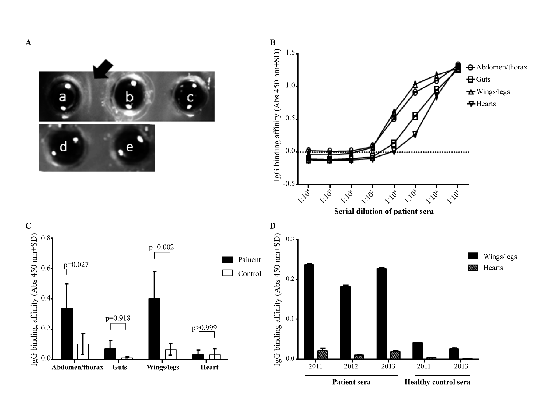
| Figure 2: Detection of anti-cricket antibodies and precipitins between patient sera and cricket antigen: A. Immunodiffusion betweenwhole cricket extracts (a) and the patient sera (b) compared with a negative control (c) and also between whole cricket extracts (d) and normal control sera (e). Arrow indicates antigen-antibody lines of precipitation. Arrow indicates antigen-antibody lines of precipitation. B. Titration of anti-cricket antibodies showing the highest titer of antibody against the wings/legs and abdominal/ thoracic wall extracts. C. Comparative binding to cricket antigens demonstrating significantly higher affinity binding of patient sera than a control subject to wings/legs and abdominal/thoracic wall proteins (n=3). D. Persistent high levels of antibodies against wings/legs of cricket in patient sera overtime. Two-way ANOVA with Sidak’s multiple comparisons tests was used for statistical analysis. |