|
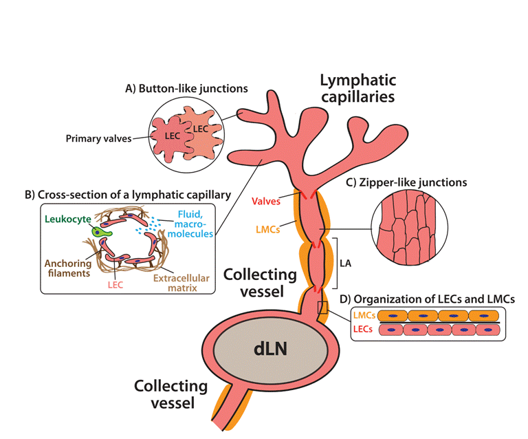
| Figure 1: Organization of the lymphatic network. (a) Afferent lymphatic vessels begin as blind-ended capillaries in peripheral tissues. These then merge into collecting vessels, which feed into a dLN. Collecting vessels contain valves and are surrounded by smooth muscle cells (SMCs). The vessel segment between two valves is called lymphangion (LA). Lymph fluid and leukocytes leave the LNs through the efferent collecting vessel. At the level of the thoracic duct the lymphcontent is ejected into the blood vascular circulation. Inserts: (A) LECs in lymphatic capillaries are oak-leaf shaped. Neighboring cells partially overlap and are connected by discontinuous, “button-like” cell-cell junctions (thick black lines). This setup generates open flaps (primary valves) (B) The flaps account for the entry of fluids, macromolecules and leukocytes into lymphatic capillaries. Capillary LECs are connected with the extracellular matrix through filaments, which account for the rapid dilation of lymphatic vessels during tissue edema. (C) LECs in lymphatic collectors have an elongated shape. Neighboring LECs are connected by continuous, “zipper-like” cell-cell junctions. This organization makes the collecting vessels less permeable as compared to initial capillaries and well suited for conducting fluids. (D) LECs in collecting vessels are in intimate association with lymphatic muscle cells (LMCs) lymphatic muscle cell, which mediate lymphatic contractility. |