|
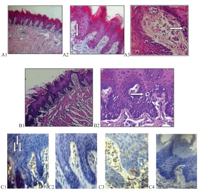
| Figure 2: Saline/chamomile extract treated group: At day 8, the dorsal surface of tongue reveled atrophy of filiform papillae, with loss of the normal appearance and most of them showed flattening of the tips with loss of their characteristic conical shape (A1, Trichrome, x100). Vacuolar degeneration was mostly seen associated with the basal epithelial layer of filiform and fungiform papillae, arrows (A2, Trichromex400; A3, H & E x400). At day 12, more changes seen in the shape of papillae and rete ridges with separation of keratin from the surface of papillae, the deeply stained nuclei, and the vacuolated cells are increased in number and seen in basal and supra basal layer, arrows (B1, H & E 100; B2, H & E x400). Mild (arrows) and negative Ki-67 immune reactivity in nuclei of epithelial cells at day 8 and 12 respectively (C1, immunohistochemistry ×400; C2 immunohistochemistry ×400). Mild and negative cytoplasmic reaction to Bcl-2 in tongue epithelium at day 8 (C3, immunohistochemistry x400) and 12 (C4, immunohistochemistry x400) respectively. |