 |
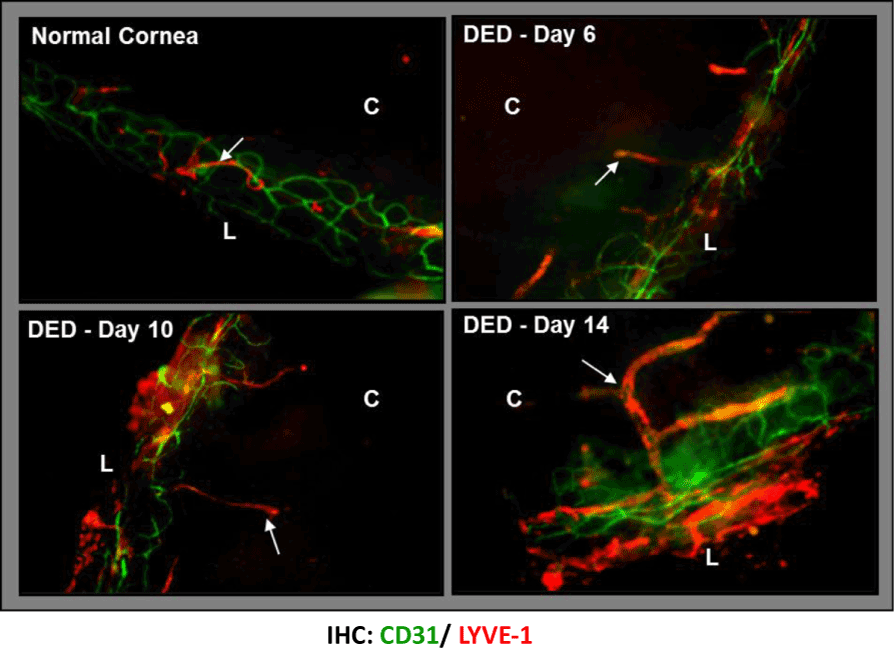
| Figure 3: Selective ingrowth of corneal lymphatic vessels in dry eye disease. Confocal micrographs showing corneal lymphangiogenesis in normal cornea and in dry eye corneas at days 6, 10, and 14 post-induction of dry eye in a mouse model of dry eye disease. The lymphatic vessels (LYVE1+CD31lo) increase both in area and caliber, and grow towards the cornea center with disease progression. The lymphatics are unaccompanied by blood vessels (CD31hi/LYVE1-). (Lymphatics marked by arrows; C: Cornea; L: Limbus). Adapted from [81]. |