|
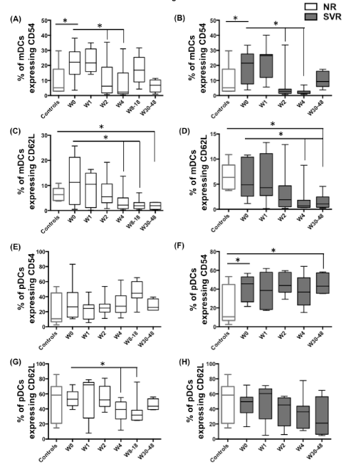
| Figure 4: IFN/RBV treatment downregulates the frequency of CD54+ mDCs, CD62L+ mDCs and CD62L+ pDCs in NRs: PBMCs isolated from seronegative controls, NRs and SVRs were stained with polychromatic antibody cocktail described in supplementary Figure 1. (A) Box and whiskers graph indicating the percentage of mDCs expressing CD54 in NRs, and (B) SVRs. (C) Box and whiskers graph indicating the percentage of mDCs expressing CD62L in NRs, and (D) SVRs. (E) Box and whiskers graph indicating the percentage of pDCs expressing CD54 in NRs, and (F) SVRs. (G) Box and whiskers graph indicating the percentage of pDCs expressing CD62L in NRs, and (H) SVRs. P values were calculated using Student’s t test (* represents P<0.05). |