|
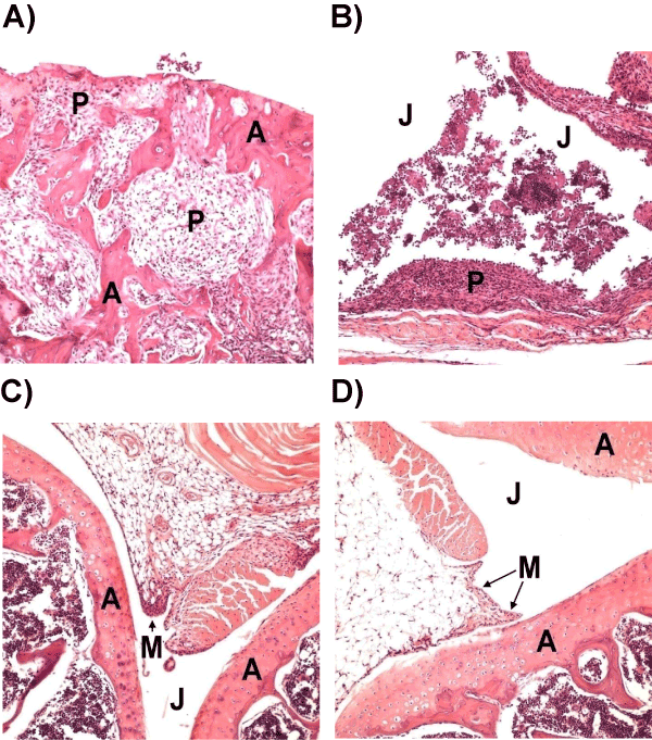
| Figure 3: Histological analysis of knee joints in peptide- and PBStreated mice. A & B, Images of the joints of PBS-treated mice with articular cartilage damage as well as a collection of cellular exudates within the joint spaces. C, Image of mouse joint treated with CII peptide, which appears essentially normal. D, Image of the joint of a mouse treated with CII-BPI-2 showing minimal inflammation. J: Joint space; M: Synovial membrane; P: Pannus formation; A: Articular cartilage. |