 |
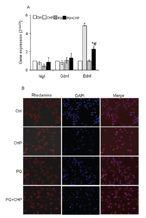
| Figure 5:Cyclo (His-Pro) induces brain-derived neurotrophic factor expression. (A) hSOD1G93A microglial cells, treated as described, were used to determine changes in gene expression after 6h PQ exposure. Gene expression values were normalised to Gapdh and presented as 2-ΔΔCt. Relative mRNA gene abundance in untreated cells was assumed to be 1.0 (control). (BDNF-F3.59= 54.01, P< 0.001 two-way ANOVA, n=3). Data represent mean ± S.D. *vs. untreated cells. #vs. PQ-treated cells. (B) CCMs from hSOD1 G93A treated for 24h with 25μM PQ, either in the presence or in the absence of 50μM cyclo (His-Pro), were added to PC12 cells for 48h. Cells were then stained with (TRITC)-labeled phalloidin and nuclei counter-stained with DAPI. Magnification 20X. The images are representative of one out of three separate experiments. |