|
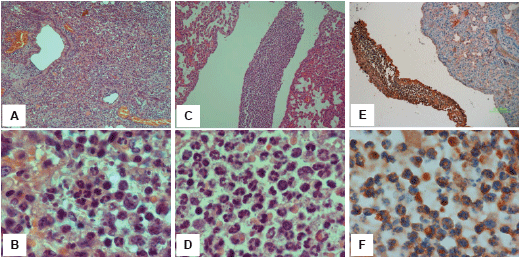
| Figure 3: Mouse model of empyema. Mice were inoculated intranasally with 2 × 10⁸ CFU of S. pneumonia (strain D39). Control mice were inoculated with equal volume of saline. Mice were sacrificed 3 days after inoculation and pleural fluid was collected and cleared by centrifugation; Lung tissue was harvested, fixed, embedded in paraffin and subjected to pathological evaluation and immunohistochemical analysis. Shown are representative H&E staining of the inflamed lung (A, B), and pleural space (C, D). Inflammatory cells in the pleural space are stained positive for heparanase (E, F). Original magnification: A, C, E x10; B, D, F x100. |