|
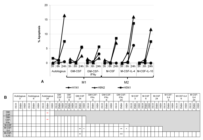
| Figure 2: Apoptosis of influenza virus-infected human M1 and M2 macrophages. (A) Differentiated macrophages mock-infected or infected by influenza viruses were fixed with 4% paraformaldehyde, permeabilized and stained for rabbit active caspase 3. The chart showed the median of three different experiments from 3 independent donors. (B) Table shows statistical significance of % of apoptosis of the different macrophages at each time point *p<0.05, **p<0.01, ***p<0.001. Red shows that the reference cell line (listed at the top of column) is significantly higher than the comparator (cells listed on the left) and black shows that it is differently lower, using two-way ANOVA followed by a Bonferroni multiple-comparison test. |