|
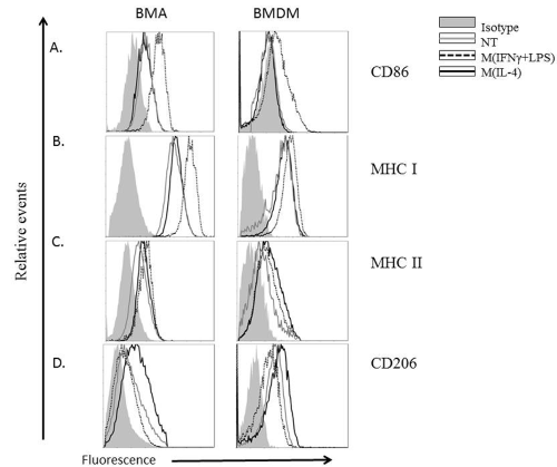
| Figure 3: Cell surface marker expression profiles for polarized macrophages. Flow cytometry analyses of the profile of BMA and BMDM treated with either IFNγ+LPS or IL-4. Changes in expression are assessed by comparison against non-treated (NT) cells. Histograms show surface staining for CD86, MHC I, MHC II, and CD206 and their differential expression in activated Mϕ. Figures are representative of one out of three independent experiments. |