|
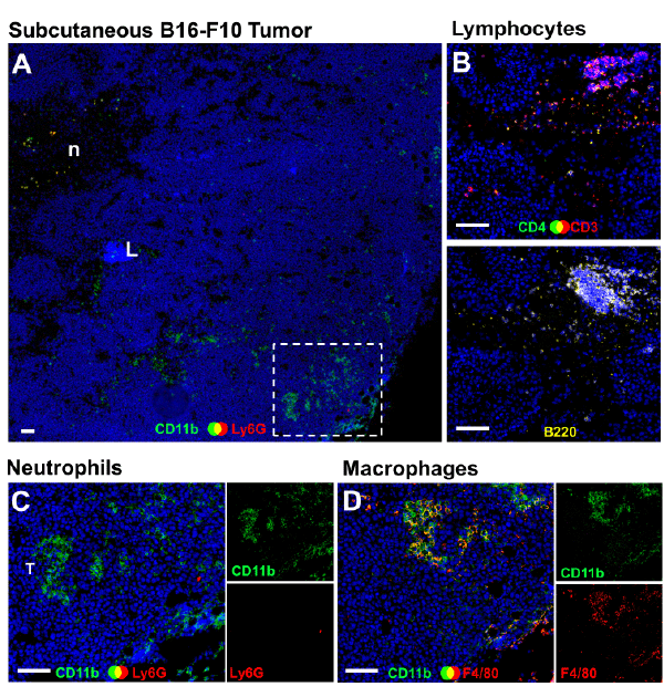
| Figure 1: Macrophage localization in subcutaneous B16-F10 murine melanoma tumours. Triple-color immunohistochemical staining ofsubcutaneous B16-F10 murine melanoma tumors shows that CD11b+ Ly6G- myeloid cells (A, green) are a localized predominantly in theperipheral parenchyma (dashed box), many of which are F4/80+ macrophages (D, yellow/orange), and not neutrophils (C, green). Serialmicrographs are representative of 3 tumors, excised 15 days after subcutaneous inoculation with 1 × 10⁶ B16-F10 cells into the left flank ofC57BL/6 mice, and processed as described previously [14,65]. ‘N’ denotes a region of central necrosis; ‘L’ denotes a dense cluster oflymphocytes deep within the tumor, that stain brightly for T-cells (B, top panel), and B-cells (B, lower panel). Side panels (C and D) showsingle color channels for merged micrographs. All sections were counterstained with DAPI (blue). White scale bars: 100 μm. |