|
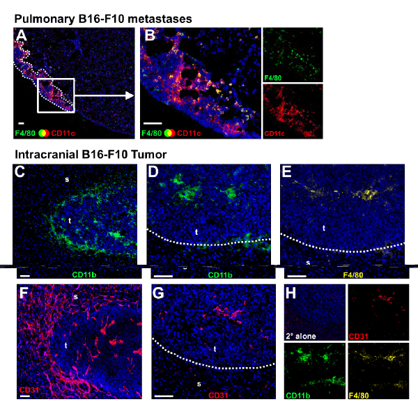
| Figure 2: Macrophage localization within pulmonary and intracranial B16-F10 tumors. Triple-color immunohistochemical staining ofpulmonary and intracranial B16-F10 tumors in C57BL/6 mice shows that macrophages are associated with growing tumors in these locations.(A, B) Interstitial or inflammatory macrophages (F4/80+ CD11c-, green) are found within pulmonary metastases while lung resident alveolarmacrophages (F4/80+ CD11c+, yellow/orange) are a localized predominantly at the interface between invading tumor and neighboring lungtissue. Similarly, (C) Resident microglia (CD11b+ F4/80low, green) are found at the interface between the expanding tumor mass (t), and thehighly vascularized (F) surrounding striatum (s). Within the tumor, CD11b+ cells (D, green) appear to be closely associated with CD31+tumor vasculature (G, red), and stain brightly for F4/80+ (E, yellow); we identify these CD11b+ F4/80+ cells as macrophages recruited from thecirculation. Serial micrographs are representative of tissues from 3 separate animals for each location. Pulmonary metastases were establishedby tail-vein inoculation of 1 × 10⁵ B16-F10 cells, while intracranial tumors were established by inoculating 5 × 10⁴ B16-F10 cells as describedpreviously [53]. Lungs and brains were excised, 21 and 7 days after inoculation, respectively, and processed as described previously [14,66]with the exception that AlexaFluor 647-conjugated secondary antibodies were used to detect F4/80-, CD11b-, and CD31-directed primaryantibodies, thus avoiding autofluorescence within these tissues. Small side panels (B and H) show single color channels for each indicatedmarker, including a secondary (2°) only panel (H). All sections were counterstained with DAPI (blue). Scale bars: 100 μm. |