|
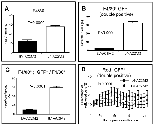
| Figure 6: Cancer cell derived IL-4 promotes phagocytic behavior in tumor associated macrophages in vivo and cultured macrophages in vitro . GPF expressing EV-AC2M2 or IL4-AC2M2 tumors were resected at 500mm3 volumes from engrafted Rag2-/- ; IL2Rγc-/- mice. Single cell suspensions were stained with PE-conjugated F4/80 antibody to identify macrophages and analyzed by flow cytometry (n=4 for each cohort). A: F4/80+ macrophages were elevated in IL4-AC2M2 relative to EV-AC2M2 tumors (P=0.0002). B: F4/80+ GFP+ double positive cells were elevated in IL4-AC2M2 relative to EV-AC2M2 tumors (P<0.0001). C: The proportion of F4/80+ cells which were double positive for F4/80 and GFP was elevated in IL4- AC2M2 relative to EV-AC2M2 tumors (P=0.0001). D: Orange cell tracker dye labeled peritoneal macrophages (red) were co-cultured with either GFP-expressing EV-AC2M2 or IL4- AC2M2 cells for 24 hours. Nine random fields were then selected from each co-culture and imaged every 5 minutes for 18 hours by video time-lapse microscopy. Numbers of yellow cells and red macrophages were quantified using ImagePro software. Higher percentages of yellow cells were found in IL4-AC2M2 co-cultures (P<0.0001; n=9 for both groups). |