|
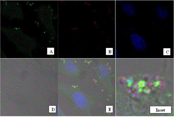
| Figure 4: Phagocytosis of Staphylococcus aureus particles. HeLa OptMR stable transfectants were incubated with approximately 5:1 ratio of pHrodo SA particles per cell for 30 minutes. Following incubation, cells were stained with monoclonal anti-MR and secondary stained with goat anti-mouse Alexa 488. Images were captured with a 63x oil objective on the Zeiss LSM-510 (A). pHrodo SA particles are shown (B). Nuclear staining was achieved with ToPro-3 (C). Brightfield view of the cells (D). Merge panel (E). Panel F shows the digital zoom of the designated inset in panel E. |