|
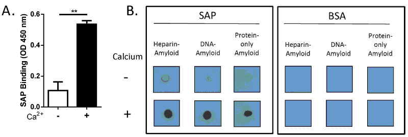
| Figure 1: SAP binding to amyloid fibrils containing cofactors. (A) Binding of SAP to Aβ (1-42) (10 μg/ml) in the absence or presence of 2 mM Ca2+ was assessed by ELISA. Error bars are means ± SEM of duplicate wells (**p<0.005 compared with no Ca2+). Similar results were obtained from five independent experiments. (B) Heparin-containing amyloid, DNA-containing amyloid, and protein-only amyloid were mixed with biotinylated SAP or biotinylated BSA in the absence or presence of 2 mM Ca2+. After several washes, the precipitates were dotted on a membrane, and binding was detected by chemiluminescence. Similar results were obtained from three independent experiments. |