 |
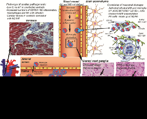
| Figure 1: M1 and M2 macrophages in HIV/SIV pathogenesis of brain, dorsal root ganglia, aorta and ventricle tissues. M1 and M2 monocytes are not well defined in blood vessels, but in tissues are defined as inflammatory M1 and anti-inflammatory M2 macrophages. In brain, the correlates of neuronal damage include: activated and infected macrophages and parenchymal microglia, M1 (MAC387)/M2 (CD163) ratio, monocyte/macrophage (MΦ) accumulation, and perivascular (PV) cuffs. In DRGs, few M1 monocytes are in uninfected DRGs. M1 macrophages correlate with severe DRG histopathology and loss of intraepidermal nerve fibers. M2 macrophages are elevated in DRGs, but do not correlate with severity of pathology. In heart, there are low to no M1 macrophages in uninfected tissue. The numbers of M1 and M2 macrophages increase with HIV infection and M2 macrophages correlates with cardiac fibrosis. Pathways of cardiac pathogenesis in aorta include the development of M2 foam cells, M1 and M2 macrophage accumulation in the intima, reduced reversed cholesterol transport and thickening of the intima-media. MNGC: Multi-Nucleated Giant Cell. |