 |
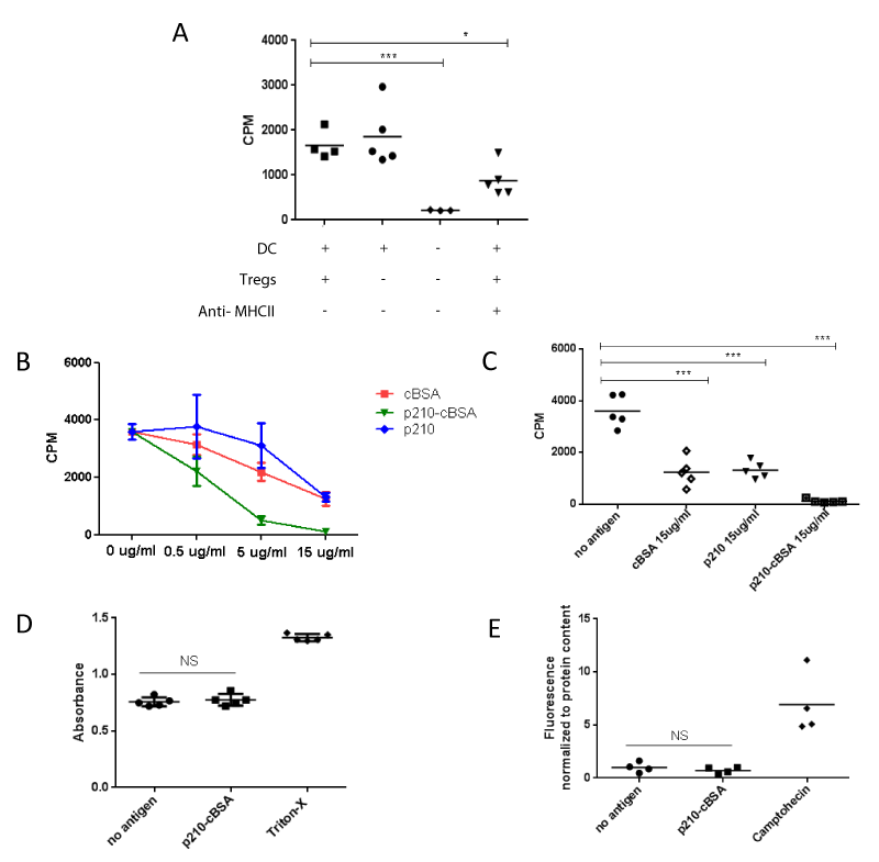
| Figure 2: p210-cBSA dose-dependently inhibits T effector cell proliferation without being cytotoxic in an in vitro model of T cell function. Proliferation of polyclonally activated T effector cells was determined in the presence or absence of DCs, Tregs and/or a neutralizing MHC class II antibody (A). Proliferation of polyclonally activated T effector cells cocultured with DCs and Tregs is determined in the presence of increasing concentrations of p210- cBSA, cBSA or p210 (B and C). The proliferative capacity of the cells is expressed as counts per minute (CPM) and cells without antigen served as control. Cytotoxicity and apoptosis were determined in p210-cBSA treated cells by assessment of LDH (D) and caspase 3 activity (E). Triton-X and camptohecin treated cells served as controls for the LDH and caspase 3 assays, respectively. (*p ≤ 0.05 ***p<0.001) |