 |
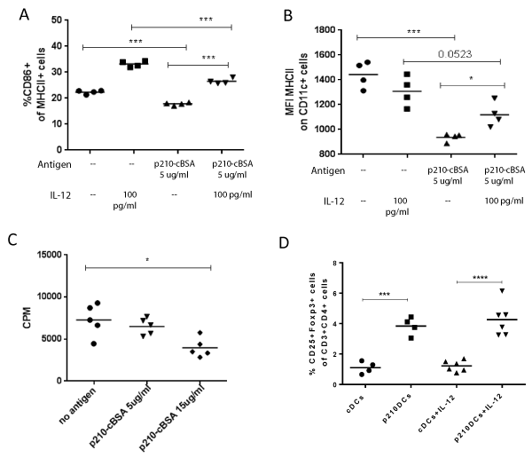
| Figure 6: p210-cBSA decrease the DC activation status and the effect on proliferation is independent of MyD88 signalling. DCs were stimulated with p210-cBSA in the presence or absence of 100 pg/ml IL-12 and the frequency of CD86+ cells out of MHCII+ cells (A) and the expression of MHC class II on all CD11c + cells (B) were thereafter determined with flow cytometry. DCs, Tregs and polyclonally T effector cells from MyD88 deficient mice were used to determine the role of MyD88 signaling in the cocultures. Proliferation was determined after 72 hours and is expressed as counts per minute (CPM) (C). The effect of IL-12 on conversion of naïve T effector cells into Tregs was studied by pulsing DCs with p210-cBSA (p210DCs) for two hours and analyzing T effector cell phenotype with flow cytometry after 72 hours coculture with and without 100 pg/ml IL-12 (D). Un-stimulated cells served as controls (cDCs) and Tregs were gated as CD25+FoxP3+ out of CD3+CD4+ cells. (*p ≤ 0.05, **p ≤ 0.01, ***p<0.001). |