|
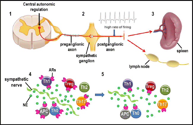
| Figure 3: Sympathetic neurotransmission in the spleen from rodent models of RA. 1, 2) Increases in the firing rate of sympathetic neurons that supply the spleen. Preganglionic sympathetic neurons stimulated by descending axons from central regulatory nuclei (e.g., the rostroventral lateral medulla) drive hyperactivity. 2, 3) The greater reactivity of the preganglionic neurons induces an increase firing rate in postganglionic sympathetic neurons (e.g., superior mesenteric-celiac ganglion) that send their axons to the spleen. Sympathetic nerves that enter the spleen (and lymph nodes) reside in T cells regions and at sites of antigen presentation and processing. 4) Increased firing rates of sympathetic nerves induce greater release of norepinephrine (NE), increasing the interaction of NE with adrenergic receptors (ARs) expressed in antigen presenting cells (APC) and Th cells. Ligand binding to ARs can cause shifts in Th cell subset balance and functions. 5) Increased nerve activity over time destroys nerves (referred to as autodestruction or “dying back”). Subsequently, sympathetic nerves respond with a sprouting response in which the nerves return, but with a different pattern of distribution in immune compartments, a process termed remodeling. The high frequency of nerve firing also increases NE concentrations/availability that induces decreases β2 -AR number and sensitivity and affects AR-mediated signal pathway activation and transduction. These events also occur in the arthritic joints (not shown). |