 |
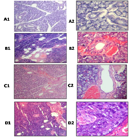
| Figure 1: A photomicrograph of submandibular salivary gland of the saline/distilled water group showing the normal architecture of salivary acini and duct (A1, H and E × 100; A2, H and E × 400). Saline/chamomile group shows normal architecture of salivary acini but with the presence of congested blood vessels. Some of the secretory elements as well as ductal elements revealed complete degeneration leaving large vacuoles (B1, H and E × 100; B2, H and E × 400). 5-FU/distilled water treated group showing loss of normal architecture of salivary acini; the acinar cells have small pyknotic nuclei and vacuolated cytoplasm (C1, H and E × 100; C2, H and E × 400). 5-FU/chamomile extract treated group showing more loss of normal architecture of salivary acini and disturbed lobular structure; the acinar cells showing vacuolated cytoplasm and contain nuclei of different sizes and shape. The secretory elements as well as ductal elements revealed complete degeneration leaving large vacuoles (D1, H and E × 100; D2, H and E × 400). |