 |
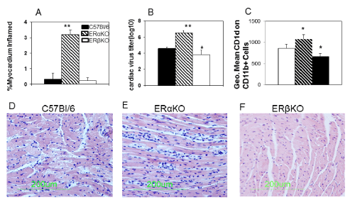
| Figure 1: ERα suppresses myocarditis susceptibility. Female C57Bl/6, ERαKO and ERβKO mice, 4-7 weeks of age, were infected i.p. with 102 PFU CVB3 and killed 7 days later. Hearts were evaluated for (Sections A, D-F) myocarditis by staining formalin fixed sections with hematoxylin and eosin. Percentage myocardium inflamed for the total number of mice/group was determined by image analysis (A). Representative section from an individual mouse/group is shown in (D-F). Virus titers in the heart were determined by the plaque forming assay (B). Spleen lymphocytes were isolated from individual infected and uninfected mice, stained for CD1d and reported as mean fluorescence intensity gating on the CD11b+ population (C). Results are given as mean ± SEM from 5-6 mice/group. * and **Significantly different than C57Bl/6 mice at p<0.05 and p<0.01, respectively. |