 |
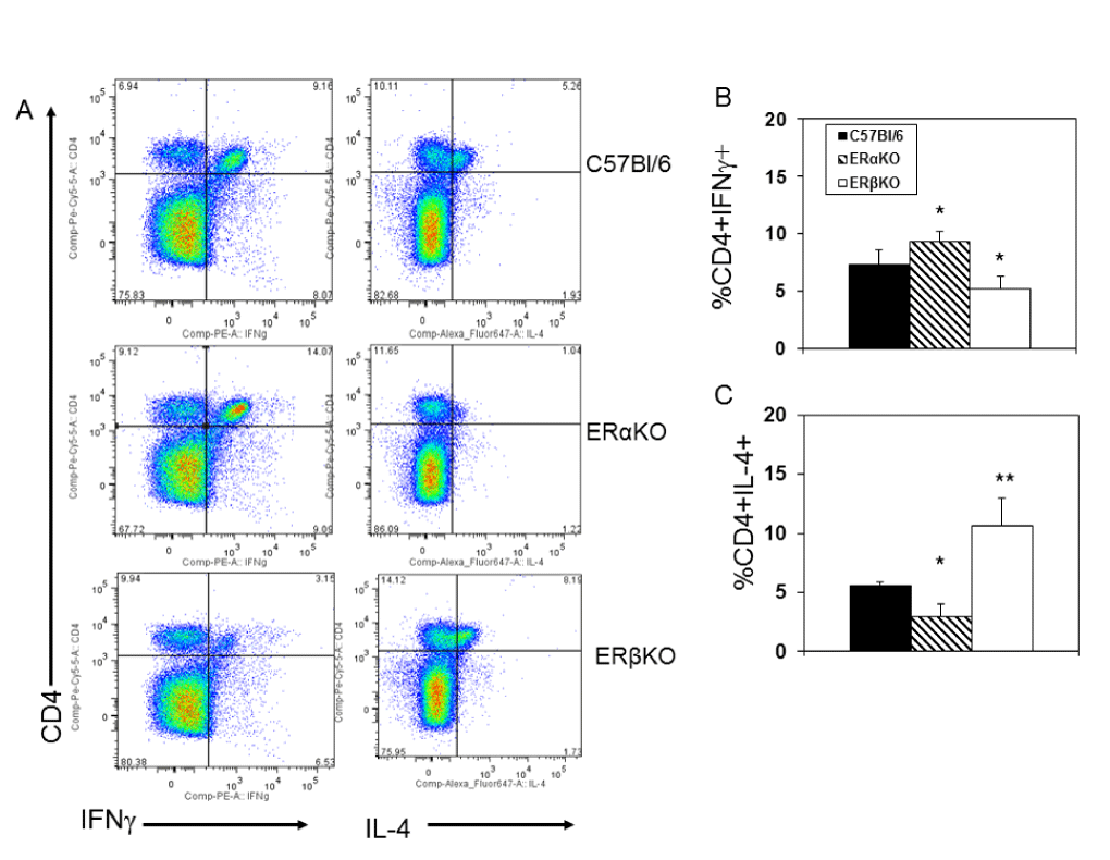
| Figure 2: ER modulates IL-4 and IFNγ expression by CD4+ T-cells. Spleen cells from C57Bl/6, ERαKO and ERβKO mice infected 7 days earlier with 102 PFU CVB3 were cultured for 4 h in medium containing PMA, ionomycin and brefeldin A, labeled with antibody to CD4, fixed, permeabilized, and labeled with antibodies to IFNγ and IL-4. Cells were evaluated by flow cytometry. (A) Representative flow diagrams from an animal in each group; (B) Mean (%) of cells staining positive for CD4 and IFNγ ± SEM. (C) Mean (%) cells staining positive for CD4 and IL-4 ± SEM. Groups consisted of 5-6 mice each. * and ** Significantly different than C57Bl/6 mice at p<0.05 and p<0.01, respectively. |