|
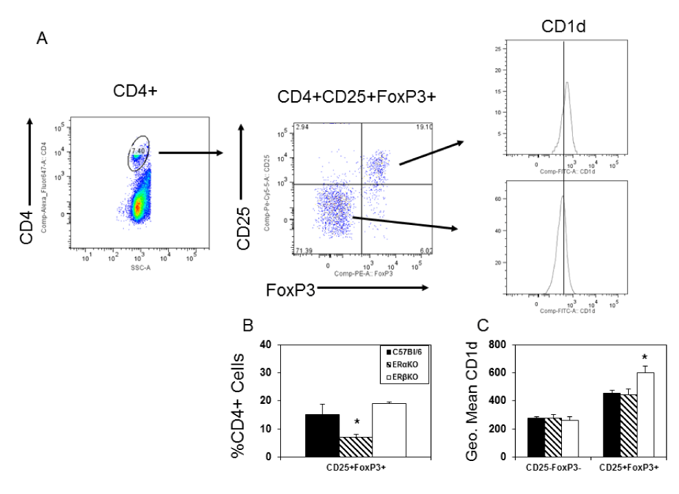
| Figure 3: ERα promotes CD4+ T regulatory cell response. Spleen cells from C57Bl/6, ERαKO and ERβKO mice infected 7 days earlier with 102 PFU CVB3 were labeled with antibodies to CD4, CD1d and CD25, fixed, permeabilized and labeled with antibody to FoxP3 for flow cytometry analysis. (A) Cells were gated on the CD4+ cell population from an infected C57Bl/6 mouse, and then evaluated for CD25 and FoxP3 expression. The CD4+CD25-FoxP3- and CD4+CD25+FoxP3+ subpopulations were evaluated for CD1d expression. The line in the CD1d graph at far right indicates mean intensity fluorescence for the CD4+CD25-FoxP3- population and indicates that the CD4+CD25+FoxP3+ cells express higher levels of CD1d. (B) Summary of percentage of CD4+ cells which are CD25+FoxP3+ for 5-6 mice/group (mean ± SEM). (C) Summary of mean fluorescence intensity of staining for CD4+ cells which are either CD25-FoxP3- or CD25+FoxP3+ for 5-6 mice/group (mean ± SEM). *Significantly different than C57Bl/6 mice at p<0.05. |