 |
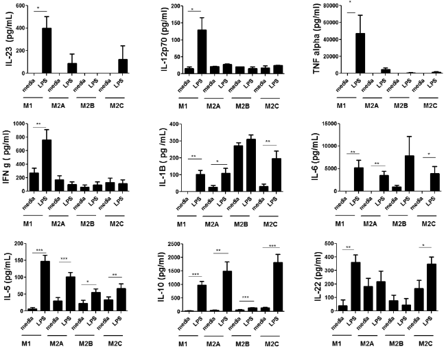
| Figure 3: Cytokine production in polarized macrophages following LPS stimulation. MDMs were polarized using appropriate stimuli for 2 days: IFNγ (20 ng/ml) for M1 macrophages, IL-4 (20 ng/ml) for M2a macrophages, LPS (1 μg/ml) and IL-1β (10 ng/ml) for M2b macrophages and IL-10 (10 ng/ml) for M2c macrophages. Polarizing stimuli were removed after the 2 days and polarized macrophages were treated with LPS (1 μg/ml) for 24 h. Cytokine levels in the supernatants were measured using flow cytometry as described in Materials and Methods. Bar graphs represent mean ± SEM, *p ≤ 0.05; **p ≤ 0.005; ***p ≤ 0.0005 with at least n=5.. |