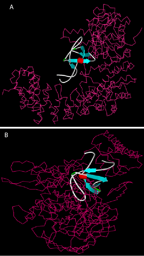
B) Mutant MEFV with PTPN12. Receptor shown with Ca wire
display style and pink color, ligand: schematic display style.
 |
| Figure 2: Visualization of docking complex. A) MEFV with PTPN12 B) Mutant MEFV with PTPN12. Receptor shown with Ca wire display style and pink color, ligand: schematic display style. |