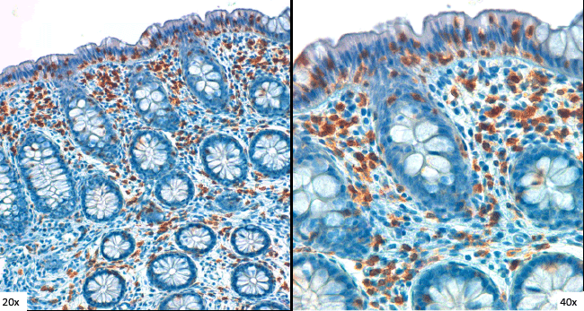
Figure 4:
Immunohistochemical analysis of T lymphocytes in LC with anti-CD3 antibody (DAKO). Colonic mucosa showing increased IELs in a patient with lymphocytic colitis. Original magnification 200x; higher magnification, on the right 400x.